


By Berger Thermal Research Ltd: www.bergerdynamics.com
Reference: Chamber-Specific Radiofrequency Lesion Dimension Estimation Using Novel Catheter-Based Tissue Interface Temperature Sensing, Jacob S. Koruth et al. JACC 2017;10 https://www.jacc.org/doi/epdf/10.1016/j.jacep.2017.08.008
(a device of Biosense Webster, a Johnson & Johnson company)
Background: RF ablation is a common therapeutic approach for treating cardiac arrhythmias. The technique consists of advancing an ablation catheter through a vein and into the heart chamber, locating the site responsible for the arrhythmia and destroying it by using a catheter whose tip electrode is brought into contact with the target site. An RF electrical current (500kHz) is applied to the heart muscle between the tip electrode and an external return patch attached to the back of the patient. The cardiac tissue is heated via resistive heating (Joule heat) and destroys a small region which is responsible for the heart rhythm disorder. The ablation catheter used in the study was an open irrigated catheter with 56 small holes. Normal saline was injected into the electrode tip, which contained 6 embedded temperature sensors, in order to cool it down (Fig.1)
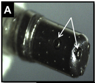
temperature sensors (white arrows)
Clinical need: lesion dimension prediction is crucial in tissue ablation to ensure effective treatment of conditions, such as atrial fibrillation by continuous monitoring of the lesion during the procedure, to achieve a durable conduction block. This could prevent complications and improve treatment outcomes.
The challenge: To develop a 3D Multiphysics model of the catheter and its interaction with the tissue that integrates electric current (Joule heat), fluid flow (blood flow and internal irrigation) and heat transfer, enabling real-time operation and closed-loop feedback using temperature measurements from sensors located at the catheter tip.
The goal of this article: COMSOL is advancing the integration of physics-based simulation with deep learning by the the surrogate model. This approach allows for real-time clinical applications, inverse problem solving, and predictive tools that were previously impossible.
The goal of the article is to demostrate the significant technology progress in the last decade by exlpaining how the problem was solved with COMSOL Multiphysics a few years ago, without these modern tools.
Model: The approach to lesion estimation was based on simulated modeling of the catheter tip during RF ablation with COMSOL. This model is a multiphysics coupled model that incorporates the following:
- the catheter tip and surrounding tissue structures
- the electrical field generated from the current source, assumed to be constant
- the electrical and thermal properties of the tissue and their relation to temperature
- the fluid dynamics of the irrigation fluid and its effect on the thermal field; and
- the lethal temperature isotherm of 57 C, which was selected to define the lesion size. To simplify the model, a single parameter of catheter penetration depth into the tissue was used to represent the possible configurations of the contact interface (“the free parameter”). The catheter itself was based on the actual structure of the study catheter, with respect to its dimensions and materials.
Build the Data Base: The main result of the simulation was the time-dependent development of the temperature field, which was used to produce the database used by the lesion assessment algorithm. A set of simulations were executed for various scenarios that spanned all combinations of ablation currents, degrees of catheter-tip penetration, and durations to generate the database. The ablations were assumed to continue from 4 to 125 s, with the current in the range 0.45 to 0.71 A, which is equivalent to 20 to 50 W, thus covering the typical ranges used clinically. The penetration depths at which the simulations were run included 0.01 to 1.5 mm for thin ventricular tissue and for thick atrial tissue, the penetration ranges being a function of both tissue thickness and mechanics (e.g., stiffness).
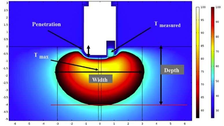
overlaid on the temperature field
The penetration depth is a parameter that represents the degree of electric and thermal contact between the catheter and the tissue, which parameterizes all unknown factors such as angle and variability in material properties. Each specific simulation run generated a temperature field around the catheter as a function of time (Fig.2). Lesion dimensions and maximum temperature (hot spot) achieved in the tissue were then defined for each simulation. Several different physical models related to the wall thicknesses were also taken into consideration.
Real time algorithm: During actual ablation, the measured generator current and maximum temperature readings were used to access this database for the relevant scenario. This selected a specific model, and through comparison of the measured temperature with the model, the lesion parameters (depth, width, and maximum temperature) were extracted and used to define lesion size. The system takes advantage of the relation between the measured temperature and the tissue-catheter interface (or penetration) for a known (measured) ablation current. For example, at a specific generator current and RF duration, the higher the measured temperature, the stronger the coupling between the tissue and catheter. The algorithm runs a few iterations to choose the best scenario with a predefined erorr between the measured value and the calculated value (finally closed the feedback loop and solved the inverse problem).
 03-9629621
  office@numerical.co.il
  נומריקל
  קבוצת קונוויזור דיגיטל